skeletal muscle tissue diagram
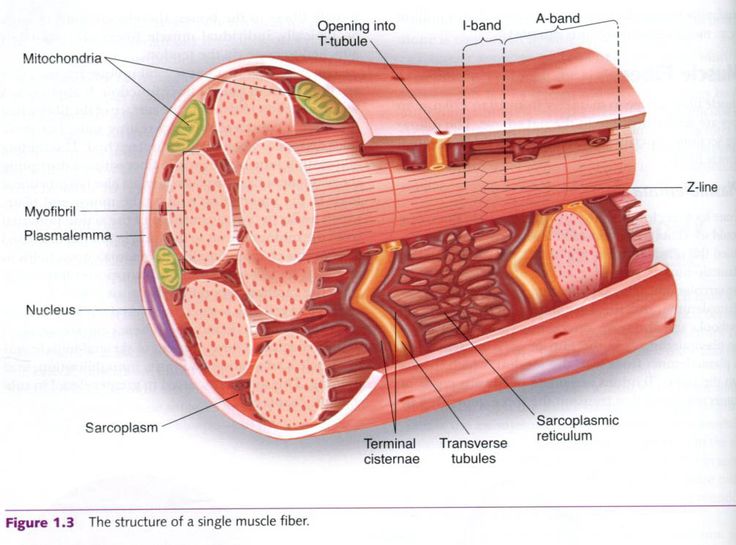
Shop as Usual Save. Sarcolemma muscle cell membrane myofibril tightly packed filament bundles found within skeletal muscle fibers M line middle of sarcomere Z line the line formed by the attachment of actin filaments between two sarcomeres of a muscle fiber in striated muscle cells H Band middle of A band.
 |
| Muscle Tissue Skeletal Muscle Smooth In A Gastrointestinal Tract And Cardiac Muscle In A Heart Types Of Mus Skeletal Muscle Smooth Muscle Tissue Cardiac |
Alternative name for skeletal muscle cells.

. In this video I have shown the simplest way of drawing Muscle drawing. Your shoulder muscles hamstring muscles and abdominal muscles are all examples of skeletal muscles. From the skeletal muscle histology slide you might identify the following important structures under the light microscope. A to Z Discovery.
Get Cash Back on Top of Your Credit Card Rewards. Epimysium perimysium and endomysium. Perimysium connective tissue layer covering the fascicles. Skeletal Muscle Tissue Diagram Quizlet Science Biology Anatomy Skeletal Muscle Tissue STUDY Learn Flashcards Write Spell Test PLAY Match Gravity Created by katrina325 Terms in this set 63 myo- latin for muscle sarco latin for flesh The property of skeletal muscle function that allows recoil after being stretched is ______.
Skeletal Muscle Description. These muscle cells are slender and long and are termed as muscle fibres. Skeletal Muscle Tissue Diagram Quizlet Skeletal Muscle Tissue STUDY Learn Write Test PLAY Match Created by armstrongsam18 Terms in this set 8 blood vessel. An individual skeletal muscle may be made up of hundreds or even thousands of muscle fibers bundled together and wrapped in a connective tissue covering.
Cross-section of skeletal muscle 3. They make up between 30 to 40 of your total body mass. Fascia connective tissue outside the epimysium surrounds and separates the muscles. Composed of all three sheaths.
The majority of the muscles in your body are skeletal muscles. Sarcoplasmic reticulum SR. Plasma membrane of the muscle cell. Tendons tough bands of connective tissue attach skeletal muscle tissue to bones throughout your body.
Please try to find out these structures from the skeletal muscle slide labeled images. Figure 1 Structure of skeletal muscle Muscle fibres are surrounded by supportive layers of connective tissue. Cordlike tendon formed from connective tissue extending beyond end of muscle fibers. Internal Structure of a Skeletal Muscle Cell Label this diagram.
Contains the genetic material. Each group of muscle fibers resembling a bunch of sticks Endomysium Fine sheath of loose connective tissue consisting mostly of reticular fibers surrounding each muscle fiber Tendon. Endomysium surrounds individual muscle fibres Perimysium surrounds a bundle of muscle fibres forming a fascicle functional unit Epimysium surrounds the entire muscle Skeletal Muscle Fibre Types. Ad Get Cash Back Rewards Exclusive Coupons from Your Favorite Stores.
Learn vocabulary terms and more with flashcards games and other study tools. The skeletal muscle fibres are multinucleated. Structure that firmly attaches muscle to bone Epimysium connective tissue layer covering the outermost part of skeletal muscle. The muscle tissue is composed of a large number of myocytes or muscle cells.
It is the pen diagram of Skeletal Smooth and Cardiac Muscle for class 10 11 and 12. Thick filaments only I band light band A band dark band H band. Protects the muscle from damage by friction carries blood vessels nerves to the muscle and helps to form the tendon. Interconnecting tubules of endoplasmic reticulum that surround each myofibril.
The culinarily appropriate components of animal meat are primarily skeletal muscle and adipose tissue 3. The muscle tissue is divided from the neighbouring. Relevant cell lines to grow these tissues would be satellite cells and adipose-derived. Ad Free 2-day Shipping On Millions of Items.
Connective tissue structure joining skeletal muscle to bone. Muscle tissue types organization biological levels cardiac smooth skeletal biology vector illustration anatomy. 9 Skeletal Muscle Tissue. Skeletal muscle fibers of the longitudinal section 3.
They are arranged parallel to each other along with some intervening connective tissue. Longitudinal section of skeletal muscle 2. Each muscle is surrounded by a connective tissue sheath called the epimysium.
 |
| Skeletal Muscle Tissue Skeletal Muscle Muscle System Muscle Tissue |
 |
| Myofibrils Complete Soccer Training Functional Anatomy Of The Skeletal Muscle Smooth Muscle Tissue Muscle Skeletal Muscle Anatomy |
 |
| Cumulative Topic 6 Microanatomy Of Myofiber Body Muscle Anatomy Muscle Anatomy Skeletal Muscle Anatomy |
 |
| Muscle Cell Types Skeletal Muscle Muscle Tissue Skeletal Muscle Anatomy |
 |
| Skeletal Muscle Muscular System Muscle Anatomy |
Posting Komentar untuk "skeletal muscle tissue diagram"